Genitourinary: Bladder Tumors

This gross photograph is of a resected bladder which has been opened to reveal the mucosal surface. There is a large invasive transitional cell carcinoma which can be recognised by its irregular, nodular, and hemorrhagic surface which contrasts with the normal smooth, glistening tan mucosa with regular folds seen in the center of the specimen.
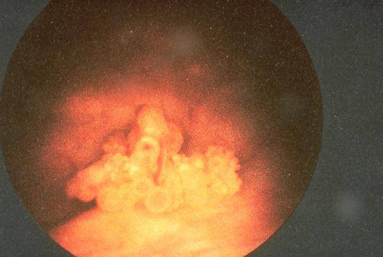
This view obtained by cystoscopic examination of the bladder shows a typical papillary transitional cell carcinoma.
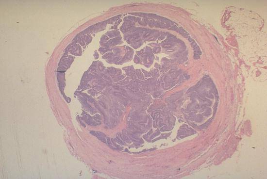
This is a low power photograph of a histologic section of a ureter with a papillary transitional cell carcinoma projecting into the lumen. There is no obvious invasion of the muscular wall of the ureter.
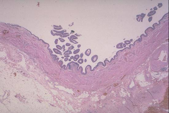
This is another section of ureter containing a non-invasive low grade papillary transitional cell carcinoma. Notice the delicate finger-like projections of the neoplasm above the mucosal surface.
This medium power photomicrograph shows the common histologic appearance of intermediate grade papillary transitional cell carcinoma. The neoplastic cells have uniform oval nuclei, abundant cytoplasm, and are arranged in ribbons of tissue supported by delicate vascular cores or "stalks".

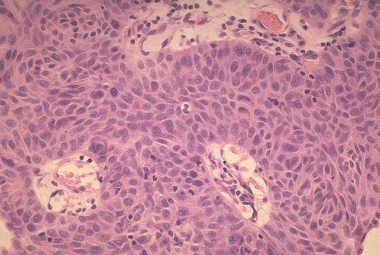
This photomicrograph shows the appearance of "carcinoma in situ" in the bladder. The epithelial cells on the left have malignant cytologic features including very large, irregularly shaped and darkly staining nuclei, which contrasts with the normal appearance of the urothelial cells on the right. Even though the abnormal cells resemble those seen in carcinoma there is no invasion of the stroma.