Pediatric Pathology: Solid Malignancies of Childhood

Kidney almost entirely replaced by fleshy tan tumor with areas of hemorrhage and necrosis. The tumor is circumscribed by a pseudocapsule. A small rim of residual kidney with attached ureter can be seen at the right.

Demonstrates pale staining stroma alternating with darkly staining blastema. Within the blastema, epithelial differentiation in the form of irregular ribbons and tubules is seen.

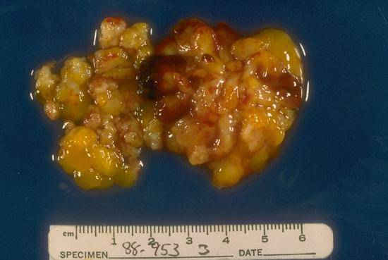
Probably arising in the adrenal, but without visible adrenal tissue in the specimen. The tumor appears lobulated and deep red to grey tan in color with extensive calcification giving it a speckled yellow appearance.

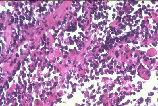
Tumor cells are small and hyperchromatic without visible cytoplasm. Pale, pink-staining neurofibrillary stroma is visible in some areas, identifying the tumor as neuroblastoma. Homer-Wright pseudorosettes are not present in this picture.
The grape-like ("botryoid") clumps of tumor are characteristic of rhabdomyosarcoma arising within any hollow structure. These tumors are associated with embryonal histology.
The microscopic features of alveolar rhabdomyosarcoma are readily apparent in this micrograph. Undifferentiated tumor cells line fibrovascular septae, with the central cells "falling out" to give the alveolar appearance. At top and right, cells with abundant eosinophilic cytoplasm and a central or somewhat eccentric nucleus demonstrate rhabdomyoblastic differentiation.