Female Reproductive Tract
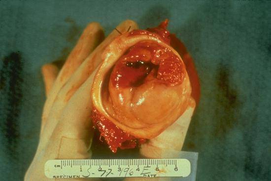
This is a specimen from a radical hysterectomy. You are looking at the cervix as you would see it on a speculum examination; note normal smooth squamous mucosa on the lower end, surrounding vaginal cuff removed with uterus and the ulcerated irregular area of the invasive carcinoma in the upper half of cervix. Cervical carcinomas can be polypoid fungating masses (exophytic) or simply an ulcerated area with predominantly inward growth into cervical stroma (endophytic).
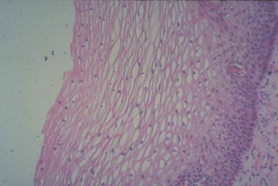
This epithelium covers the ectocervix or "portia" continuous with the vagina. It is thick during child bearing years or in response to estrogen stimulation. Note the orderly maturation of cells with nuclei becoming progressively smaller toward surface. Spaces around nuclei is the normal cytoplasmic glycogen "washed out" during processing of tissue.
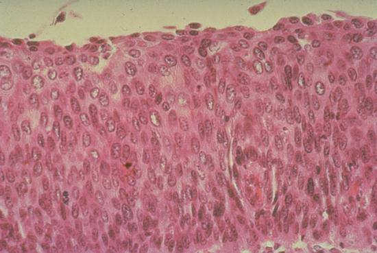
Synonymous with "severe dysplasia" which is essentially indistinguishable from carcinoma in situ (C.I.S.). Note the full thickness of the squamous epithelium has been replaced by immature cells with enlarged mildly hyperchromatic cells (slightly coarse chromatin) -- proliferating in a disorderly fashion. You may not necessarily see mitoses. This could also be called a poorly differentiated intraepithelial squamous carcinoma.

Note irregular polypoid endometrial lining (it is normally smooth). The myometrium and cervix is normal. This carcinoma was confined to the endometrial lining i.e., did not invade the myometrium; prognosis is therefore good for a cure.
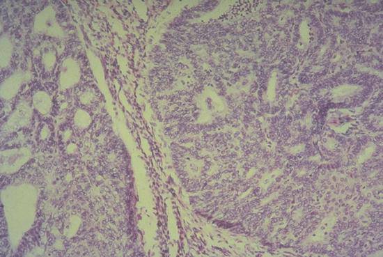
Note complex gland-forming sheets of epithelial cells forming nodular masses with thin strands of myometrial smooth muscle between. Only part of the epithelium forms glands with "solid" sheets between; this indicates a less well differentiated carcinoma.

Leiomyomas are firm white tumors with whorled patterns on their cut surfaces, such as the leiomyoma arising in the fundus of this uterus. They consist of bundles of smooth muscle surrounded by dense collagenous connective tissue.
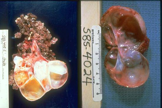
Benign cysts (right) have smooth linings. Malignant cysts (left) have epithelial proliferations that form tufted papillations. Initally, the papillations are internal but neoplastic papillations may also form on the external surface. These tumors are subclassified as serous or mucinous based on the microscopic appearance of the lining cells.
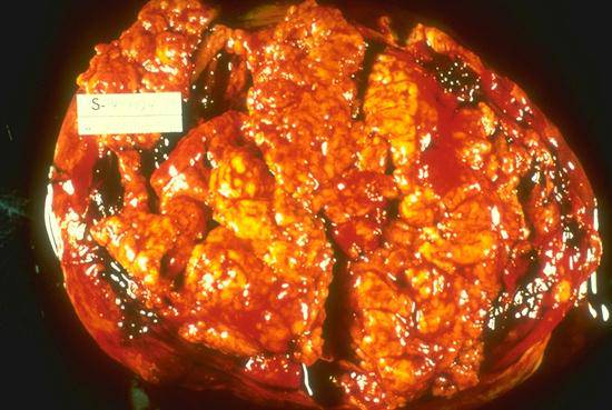
This is the cut surface of a large (about 15 cm diameter) mostly "solid" ovarian carcinoma indicating that it is high grade or poorly differentiated. The yellowish areas indicate necrosis. Lower grade carcinomas are usually predominantly cystic masses.
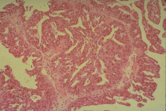
Note the complex papillary glandular architecture with the epithelial cells stratified on a supporting fibrovascular stroma. The papillary serous pattern mimics the Mullerian epithelial lining of the fallopian tube.
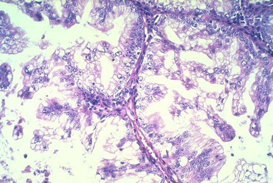
Mucinous carcinomas also form complex papillations but the tumor cells are tall, columnar, mucin-secreting cells.