Immunocytochemistry
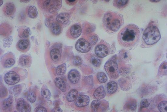
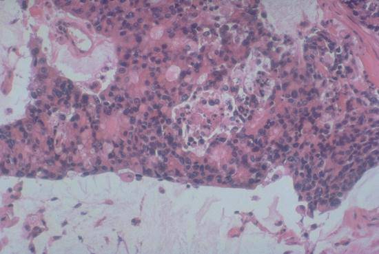
Hematoxylin and eosin (H&E)-stained section of a poorly differentiated tumor found in brain biopsy. The polygonal shape of the cells and their cohesion suggest an epithelial tumor (i.e. a carcinoma).
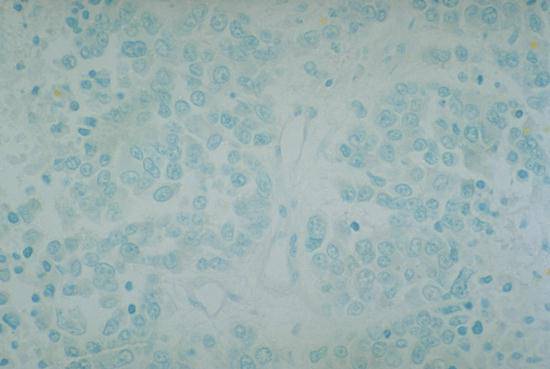
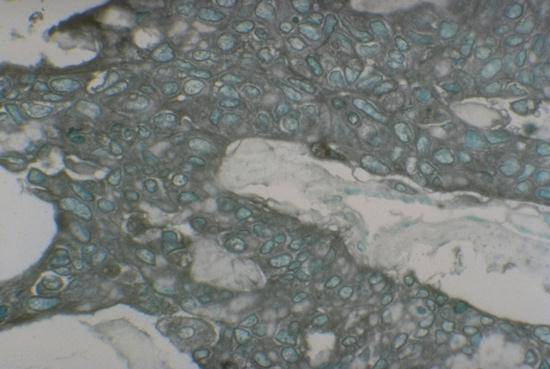
Immunocytochemical study of same tumor as in slide 1 using monoclonal antibody to low molecular weight cytokeratin using antibody 35ßH11, showing a complete absence of reactivity.
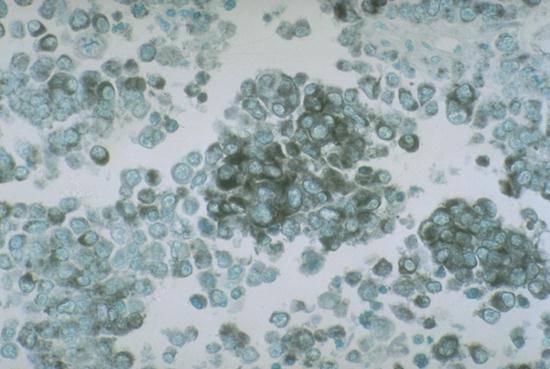
Immunocytochemical study of same tumor as in slide 1 using monoclonal antibody HMB45 to melanoma. Positive antibody binding is indicated by the black reaction product. This result indicates that the tumor is a variant of a melanoma, not a carcinoma.
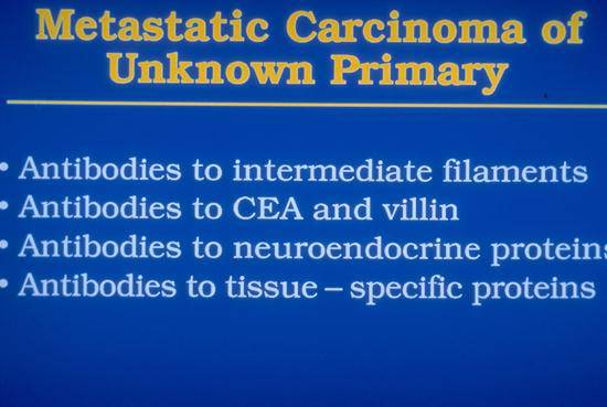
Summary of antibody studies useful in determining site of origin of carcinomas presenting at metastatic sites.
Hematoxylin and eosin (H&E)-stained section of tumor in supraclavicular lymph node found in 72 year old male. It is not obvious where this poorly differentiated carcinoma metastasized from.
Immunocytochemical study of same tumor cells as in slide 5 employing antibodies to prostatic specific antigen, showing positive reaction, and pointing to site of origin of tumor in prostate.