Infections
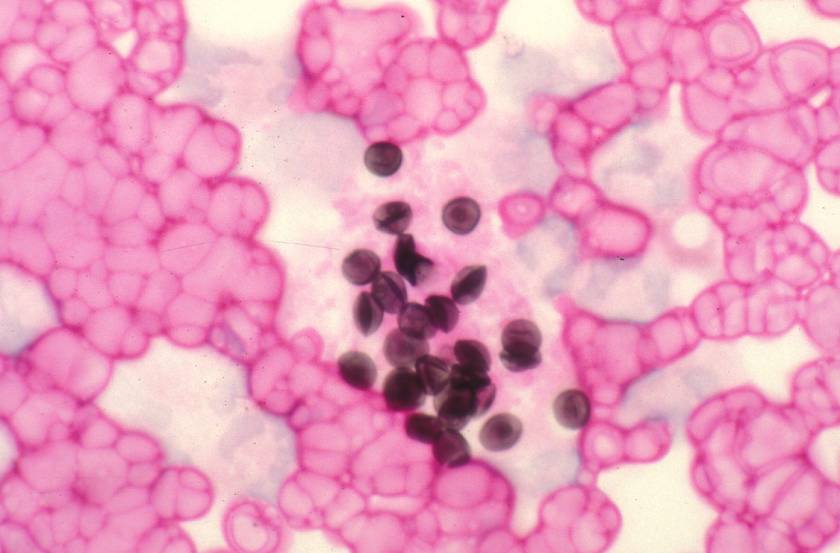
The spherical and crescentic bodies displaying the dark line or dot are the cysts of Pneumocystis carinii. Although long regarded as a protozoan parasite, it has been reclassified as a fungus on the basis of nucleic acid testing. It is the most common serious infection in U.S. patients with AIDS.
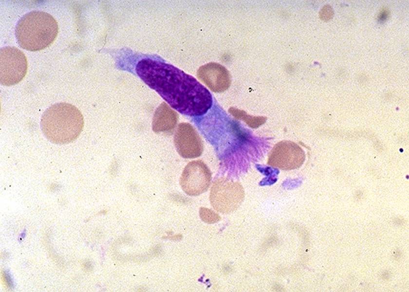
Two Toxoplasma gondii tachyzoites are lying near the ciliated end of a bronchial epithelial cell. These protozoan parasites can infect any cell in the body except the non-nucleated erythrocytes. Approximately 20% of the U.S. population is chronically infected with toxoplasma but asymptomatic. If we become severely immunodeficient, the parasites can reactivate, disseminate, and kill.
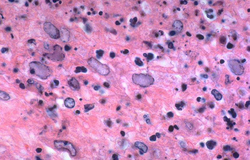
This brain biopsy displays Herpes simplex infection. The 7 or 8 infected cells show "ground glass" or "slate-gray" nuclear staining as well as thickening of their nuclear membranes as a result of chromatin margination. Many of the other cells are at different infection stages without inclusions. The viral changes shown here could also be caused by another human herpesvirus, Varicella-zoster virus (VZV).
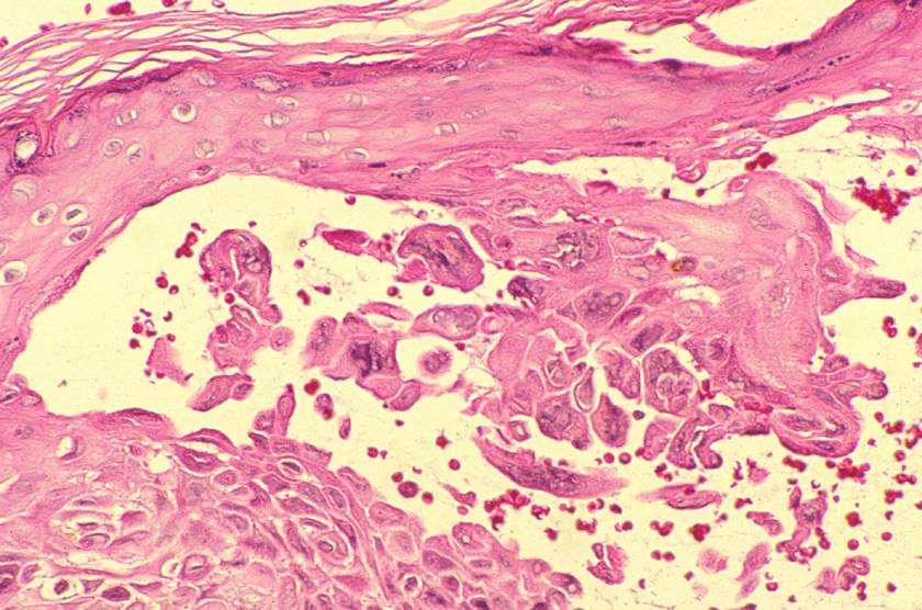
This skin vesicle has developed as a result of Varicella-zoster virus infection. The infected epidermal cells display intranuclear inclusions. Many are multinucleated. VZV and HSV cannot be distinguished from one another by classical histological staining such as hematoxylin & eosin. Immunohistological staining with virus-specific antibodies distinguishes them very clearly from one another. Specific identification is important, since treatment may vary.
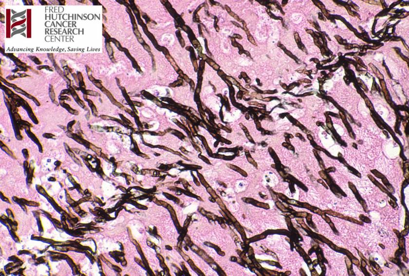
These are the fungal hyphae of Aspergillus, but their morphology does not distinguish them from other molds such as Fusarium or Pseudallescheria boydii. All three, as well as others, are characterized by these arborizing, septate hyphae which have parallel sides and usually branch at acute, equal angles -- Y-shaped or dichotomous branching.
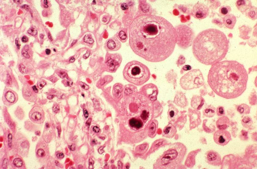
The two large cells with dark, round to oval, intranuclear inclusions surrounded by a clear halo are infected. Immediately below them is a large cell which is also infected with CMV and displays the coarsely granular cytoplasmic inclusions characteristic for this virus. Of the 8 human herpesviruses, only CMV produces both nuclear and cytoplasmic inclusions.