Male Genitourinary Pathology: Prostate
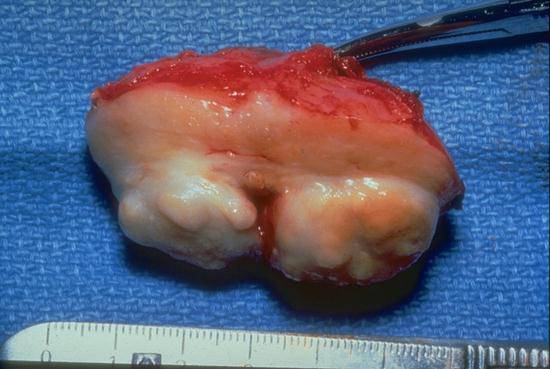
This prostate gland has been sectioned perpendicular to the urethra to reveal two large bulging masses of benign hyperplasia, one on either side of the urethra, which project above the cut surface of the surrounding tissue. This is the typical appearance of benign hyperplasia causing bladder outlet obstruction. Note how these bulging nodules can compress the urethra.

This gross photograph is of a slice of resected prostate gland which has been sectioned perpendicular to the urethra. The triangular hole in the center of the specimen is the urethra and the ruler is adjacent to the rectal surface of the gland. This gland contains carcinoma which can be seen as a yellowish mass in the lower right part of the gland (between about "8" and "10" on the ruler). Generally carcinomas arise in the periphery of the prostate, far from the urethra, where they can be felt by the examining finger.
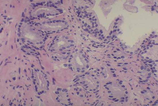
This photomicrograph shows the appearance of a low grade carcinoma of the prostate (contrast with the normal large prostatic duct in the top right corner). The glands are tightly packed together, but it is difficult to recognize definite invasive growth at this magnification. This appearance can be difficult to recognize as cancer, particularly in small needle biopsies.
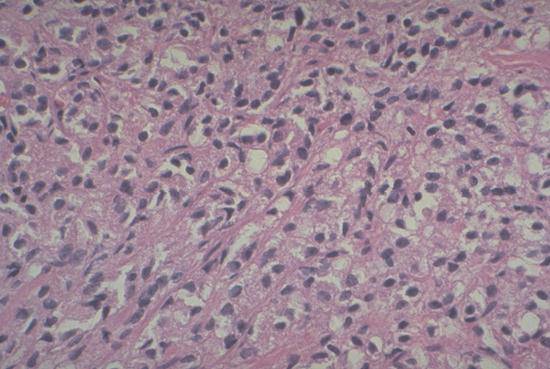
This photomicrograph shows a very high grade prostate carcinoma. The malignant cells are diffusely infiltrating the muscular stroma of the prostate, without definite gland formation. This highly invasive appearance correlates with aggressive behavior of the cancer and a high risk of metastasis.
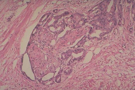
Perineural invasion is very common in prostate carcinoma, frequently involving the numerous autonomic nerves which run in the capsule of the prostate. The nerve in this picture contains a cluster of nerve cell bodies and a wavy collection of nerve fibers; the carcinoma forms malignant glands which invade into the sheath of the nerve.