Male Genitourinary Pathology: Testis

This photograph shows a bisected testis with attached epididymis and spermatic cord. The testis is the oval brown structure next to the ruler. Just below the testis and to the left is a dilated and scarred epididymis containing gelatinous brown and yellowish debris, the result of chronic epididymitis.
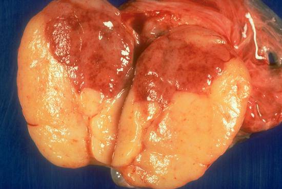
This testis has been completely replaced by a seminoma. It has been bisected to reveal the typical soft, tan, fleshy, and lobular appearance of the tumor. The area of tumor with darker coloration contains congested blood vessels.

There is only a slight amount of normal testis remaining in this bisected specimen, forming a rim of tan or brown tissue around the large, soft, ragged, hemorrhagic mass, which is an embryonal carcinoma.

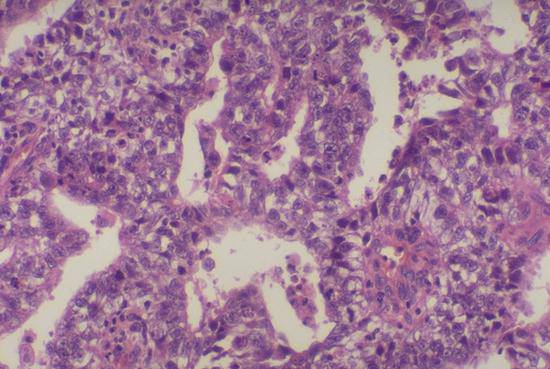
This slide shows the typical histologic appearance seminoma. Seminoma is composed of cells with abundant clear cytoplasm, central oval nuclei, and prominent nucleoli growing in sheets or in a delicate lobular pattern. There is frequently an infiltrate of lymphocytes in seminomas, as in this case. The cells in seminomas resemble to a certain extent the normal spermatogonia.
Embryonal carcinoma is composed of larger more pleomorphic (variable appearing) cells with dense cytoplasm, very large oval or irregularly shaped nuclei, prominent nucleoli, and clumped chromatin. The cells of embryonal carcinoma often form primitive epithelial structures surrounding clefts and spaces.
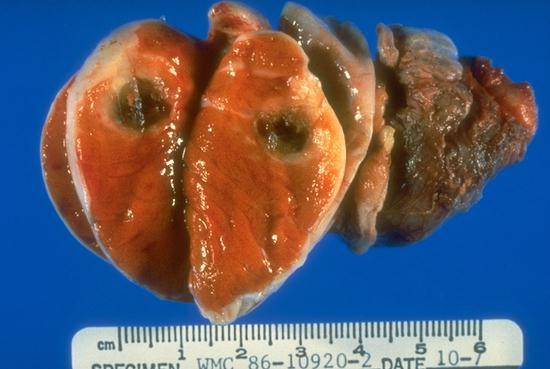
This testis has been sectioned to reveal a small, dark red (hemorrhagic) tumor mass, which proved to be a choriocarcinoma.
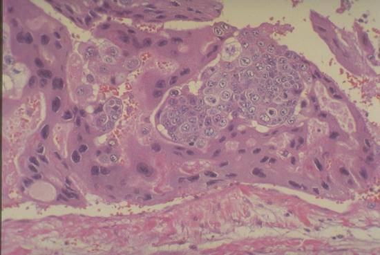
This photomicrograph shows the typical histology of a choriocarcinoma. The neoplasm resembles normal placenta with large multi-nucleated syncytial cells intermingled with clusters of polygonal cells resembling cytotrophoblasts. One huge syncytial cell weaves its way through the tissue across the entire field of this photograph. These tumors readily invade blood vessels, mimicking the behavior of normal placental cells when they implant in the uterus and tap into the maternal blood supply.