Pediatric Pathology: Neonatal
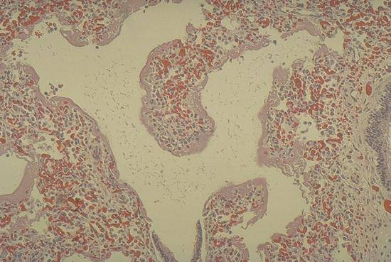
Hyaline membranes line the respiratory bronchiole and alveolar ducts of this premature baby's lung. The alveoli are atelectatic (collapsed).
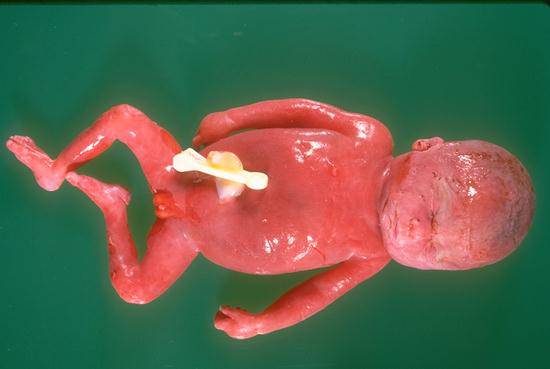
(therapeutically aborted at 23 weeks): Prenatal ultrasound examination revealed generalized edema, ascites, and pleural effusions with dilated cardiac chambers. Serological studies showed high titer of anti-toxoplasmosis IgM antibodies in the maternal circulation. This fetus is moderately hydropic as evidenced by abdominal distension and soft tissue edema.
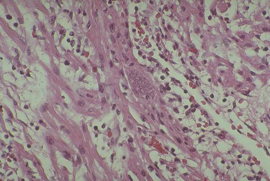
A microscopic section from the heart of the fetus shown in the preceding slide demonstrates a lymphocyte-rich inflammatory infiltrates surrounding a cardiac muscle fibers and a single large endocyst (center).
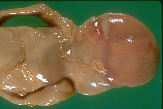
The redundant skin on the posterior aspect of the neck is supported by fluid-filled cystic spaces. Large cystic hygromas like this are often associated with generalized hydrops and/or chromosomal anomalies. This fetus was 45 XO (monosomy X, Turner's syndrome).
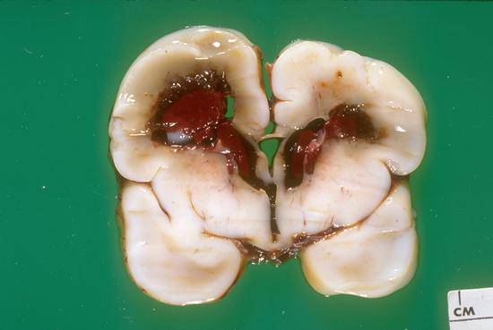
An acute consequence of generalized hypoxia is illustrated in this photograph. A large hematoma is present in the periventricular neural tissue and lateral ventricles. This type of hemorrhage usually begins in the subependymal germinal matrix adjacent to the lateral ventricle and may remain localized or may radiate into the ventricle.
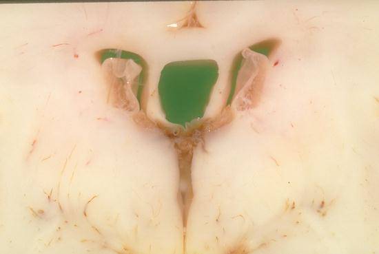
(34 week premature infant, lived for several months). If a fetus survives perinatal hypoxia and an acute periventricular hemorrhage, periventricular hematomas eventually are removed by the microglia in the surrounding neural tissue. Periventricular cysts, like those shown in this slide demarcate the locations previously occupied by blood clots. These cysts are permanent sequelae and often are associated with chronic neurological sequelae.