Respiratory: Pneumoconiosis

This composite slide illustrates the favored site of accumulation of dusts; i.e. around respiratory bronchioles. Dusts accumulate at this site as the result of local deposition and redistribution of dusts deposited in alveoli. (Photo taken from Pulmonary Pathophysiology - the Essentials, West JB, Williams and Wilkens, 1977).
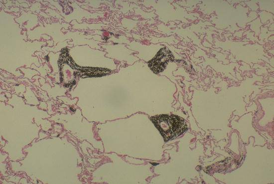
This medium-power micrograph illustrates a macule, which is simply a prominent accumulation of dust around a respiratory bronchiole. Macules are characteristic of siderosis, as in this case, but are also seen in coal-workers pnemoconiosis and in other pneumoconioses.
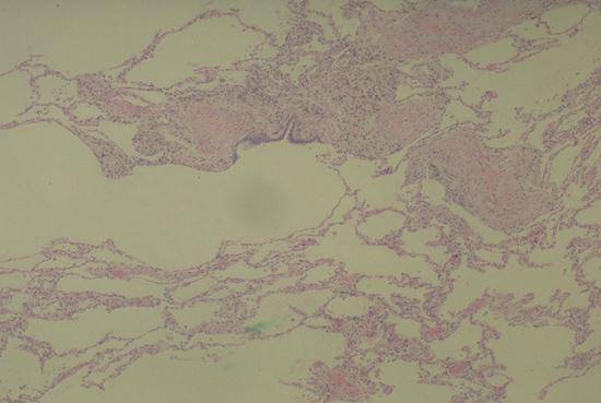
Peribronchial (peribronchiolar) fibrosis is illustrated in this figure. Fibrosis in this region is a response to heavy dust deposition and is relatively independent of the type of dust deposited. It can be seen in silicosis (this case), asbestosis, and many other disorders.

Nodular fibrosis, seen here, is a characteristic response to crystalline silica dust. The nodules seem to arise in areas of peribronchial fibrosis and can grow large enough to be easily seen on chest roentgenograms. In severe silicosis the nodules can merge to form large conglomerates that distort the lung parenchyma and cause restrictive lung disease ("complicated" silicosis).
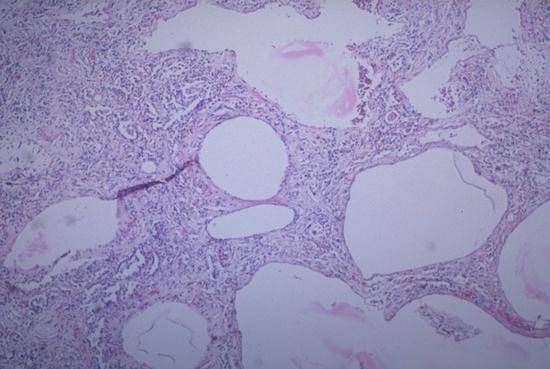
Diffuse interstitial fibrosis is a more common pattern of fibrosis. In diffuse fibrosis, collagen deposition is spread throughout the lung parenchyma rather than being restricted to peribronchial locations. In this case the diffuse fibrosis was caused by inhalation of asbestos fibers ("asbestosis") but similar alterations can also be seen in response to many other dusts.
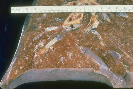
These slides contrast the gross appearance of normal lung (RS 22) with that of lung distorted by diffuse interstitial fibrosis (RS 23). One can appreciate the interlacing bands of gray-white fibrous tissue that dissect and distort the fibrotic lung. The abnormally large airspaces result from retraction of the bands of scar tissue. The pattern of enlarged distorted airspaces surrounded by bands of fibrous tissue is called "honeycombing" and is the final result of diffuse fibrosis from any cause.
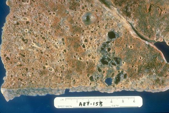
These slides contrast the gross appearance of normal lung (RS 22) with that of lung distorted by diffuse interstitial fibrosis (RS 23). One can appreciate the interlacing bands of gray-white fibrous tissue that dissect and distort the fibrotic lung. The abnormally large airspaces result from retraction of the bands of scar tissue. The pattern of enlarged distorted airspaces surrounded by bands of fibrous tissue is called "honeycombing" and is the final result of diffuse fibrosis from any cause.