Department of Laboratory Medicine & Pathology, UW Medicine, University of Washington
Submit
Search
Toggle Search Input
Toggle navigation
Menu
Submit
Search
About
Home
Message From the Chair
About Us
History, Mission, and Core Values
Justice, Equity, Diversity, and Inclusion (JEDI)
Make a Gift
Jobs
Faculty & Staff
Faculty Directory
Staff Directory
Resources for Faculty & Staff
Diagnostic Services
Diagnostic Testing
Blood Draw Locations
Anatomic Pathology Service Request
Laboratory Medicine Service Request
Anatomic Pathology Licenses & Accreditation
Clinical Pathology Licenses & Accreditation
Anatomic Pathology Research Services
Laboratory Medicine Research Testing Services
Client & Patient Services
Payment Info
Pay Your Laboratory Medicine Bill
Test Guide
Find a Laboratory Test
Specimen Collection
Critical Values
Notifiable Conditions
Staff Only Test Guide
Genetics & Solid Tumors
Molecular Hematopathology
Flow Cytometry
Immunology
Molecular Microbiology
Red Cell Disorders
Virology
Education
Graduate Program (M3D)
Medical Student Training
Residency Program
Clinical Fellowships
Research Fellowships
Post-Sophomore Fellowship
Residency & Fellowship Alumni
MS in Laboratory Medicine
MLS Undergraduate Program
MD/PhD Program (MSTP)
Certificate Program in Clinical Laboratory Leadership & Management
Training Grant Programs
Visitor Programs
Course Catalogs
Research
Research in Lab Med & Pathology
Research Testing Service (RTS)
Research Services from Anatomic Pathology Labs
Research Centers
Research Labs
DLMP Awards
Research Committee
DLMP Research Flow Cytometry Core
DLMP Research Faculty & Staff Resources
Events
Calendar of Events
Laboratory Medicine Grand Rounds
Pathology Grand Rounds
Lab Med & Pathology Research & Discovery Seminar
Lab Med Clinical Research Conference
Medical Conferences
News
Latest News
News in Patient Care
News in Education
News in Research
Contact
Resources
Toggle Page Navigation
View pages within this section
Educational Resources
Images in Pathology
Breast Lesions
Female Reproductive Tract
Gastrointestinal: Non-neoplastic
Gastrointestinal: Gall Bladder/Bile Ducts/Pancreas
Gastrointestinal: Colorectal
Genitourinary: Bladder Tumors
Immunocytochemistry
Infections
Male Genitourinary: Prostate
Male Genitourinary: Testis
Pediatric Pathology: Childhood Malignancies
Pediatric Pathology: Neonatal
Renal Tumors
Respiratory: Lung Cancers
Respiratory: Pneumoconiosis
Respiratory: URT Tumors
Sarcomas
Vasculitis
Barrett's Esophagus
Cytogenetics Gallery
Renal Tumors
US 6
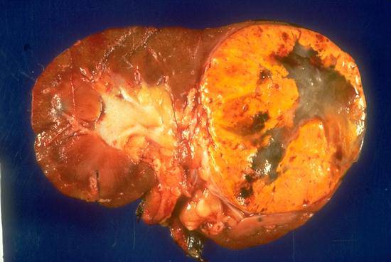
This gross photograph shows the cut surface of a kidney which has been longitudinally bisected. There is a large renal cell carcinoma in the upper pole with a typical variegated appearance with bright yellow areas, areas of hemorrhage, and tan and white areas. The bright yellow color is related to the lipid content in these tumors.
US 7
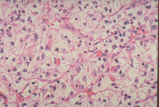
The typical "clear cell" appearance of many renal cell carcinomas is illustrated in this photomicrograph. The malignant cells have abundant clear or empty appearing cytoplasm, and the delicate lobular growth pattern is a result of the numerous capillaries between clusters of cancer cells.
$( document ).ready(function() { });