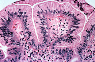
Images of Barrett’s esophagus with Low-grade Dysplasia
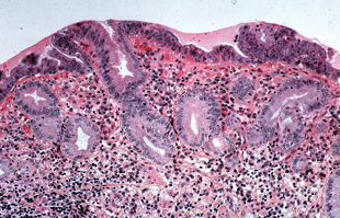
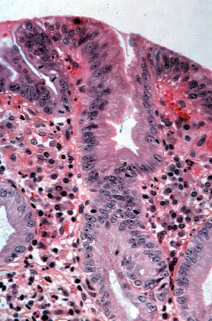
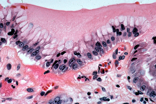
In low grade dysplasia, epithelial abnormalities extend onto the mucosal surface. The nuclei appear hyperchromatic, elongated, stratified, crowded, and may display loss of mucin production.
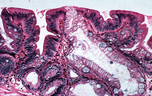
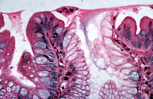
Low grade dysplasia is characterized by cells with enlarged, hyperchromatic, elongated, stratified nuclei that extend onto the mucosal surface. Typically there is decrease in the amount of mucin present - a feature which can best be appreciated at low power.
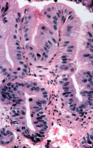
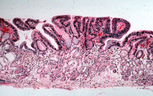
Loss of goblet cells can best be seen at low power in dysplastic areas.
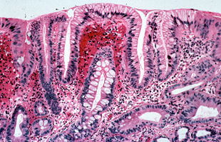
In dysplasia, the nuclear atypia extends onto the mucosal surface. Compare a biopsy with no evidence of dysplasia (on the left) with one that we felt represented low grade dysplasia (on the right).
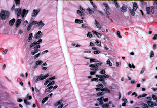
Other phenotypes of dysplasia in Barrett's esophagus
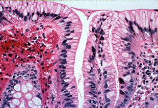
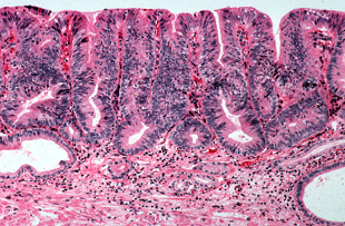
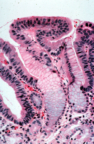
While it is more common to see mucosal surface involvement in combination with atypical deeper glands, situations may arise where only the mucosal surface demonstrates nuclear atypia while the deeper glands appear normal.
The dysplastic epithelium is present only on the mucosal surface with normal underlying glands.
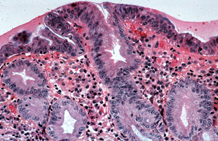
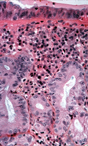
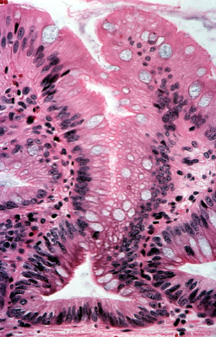
A difficult phenotype of dysplasia to recognize occurs when there is retention of apical mucin and basally oriented nuclei. At low power, the biopsy may be called negative; however, at higher power, obvious atypical nuclei can be demonstrated.
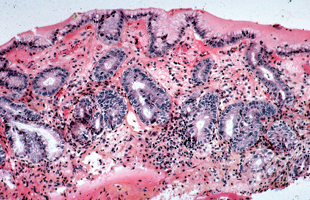
The abundant mucin seen at low power may cause elicit a diagnosis of negative for dysplasia.
At higher power, however, obviously atypical nuclei can be seen at the mucosal surface, warranting a diagnosis of low grade dysplasia.
Additional examples we would interpret as low-grade dysplasia.