Breast Pathology: Consultation Services
The breast pathology service at UW Medical Center - Montlake has expertise in the diagnosis of diseases of the breast including:
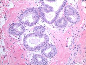
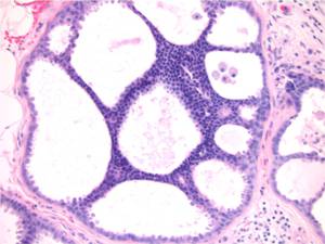
Risk Lesions
Usual ductal hyperplasia, complex sclerosing lesions, papillary lesions, atypical ductal hyperplasia, flat epithelial atypia, atypical lobular hyperplasia/lobular carcinoma in situ, etc.
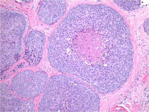
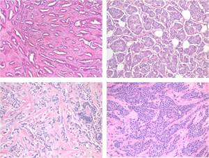
Cancers
Spectrum of invasive carcinomas and in situ carcinomas.
Fibroepithelial and Stromal Lesions
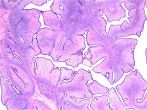
Fibroadenomas, phyllodes, pseudo-angiomatous stromal hyperplasia, fibromatosis, etc.
Special Studies
Performance and expert interpretation of immunohistochemistry and fluorescence in situ hybridization pertaining to breast pathology.
Special Studies Available
Our standard invasive breast cancer panel includes:
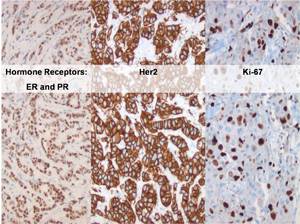
Estrogen and progesterone receptors
- Reported as an Allred score (the only clinically validated scoring system)
- >1% of cells (Allred score > 3 of 8) staining as threshold for a positive result
- Comments on status of internal controls in cases with < 1% staining
Her2/neu
- Reported and performed according to CAP/ASCO guidelines
- Our laboratory's 2007 concordance rate for Her2 IHC and FISH was > 99%
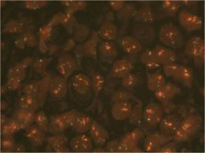
Fluorescence in situ hybridization for amplification status of HER2 gene
- Routinely performed for all cases that are equivocal by immunohistochemistry
- Reported and performed according to CAP/ASCO guidelines
- Report includes digital image
Ki-67 (MIB-1) Proliferative Index
- Provides a correlation with likelihood of response to chemotherapy
Additional Tests
Additional immunohistochemistry often performed in work-up of breast pathology cases includes (but is not limited to):
- Determination of invasive verses in situ carcinomas: Smooth muscle myosin heavy chain, p63, calponin
- E-cadherin to aid in the distinction of lobular verses ductal neoplastic processes
- Pancytokertin for identification of isolated tumor cells in sentinel lymph nodes
- Markers of basal-like subtype of invasive carcinoma: CK5/6, EGFR, p63, etc.
