Cytogenetics and Genomics Laboratories
Genetics is a branch of biology that studies the science of genes and heredity. Genes correspond to regions of the genome, which is made up of DNA. DNA is packaged in small organelles called chromosomes for proper segregation of genetic material. Cytogenetics refers to the microscopic analysis of chromosomes in individual cells. Genomics refers to the detailed molecular analysis of the entire genome. Cytogenetics and genomics studies can be performed on fresh blood, bone marrow, prenatal specimens, and solid tissue specimens, and on fixed specimens. A chromosome and/or genome analysis is considered an essential component of the important work-up for individuals with congenital malformations, mental retardation, multiple spontaneous miscarriages, or infertility, and for individuals with hematologic neoplasms and solid tumors. Information about genetic abnormalities in cancer patients can be particularly useful for establishing a diagnosis, for disease classification and monitoring, as well as for treatment decisions. Below are examples of the main types of analyses performed in the laboratory: G-banding karyotype analysis, interphase fluorescence in situ hybridization (IFISH) analysis, and chromosome microarray analysis (CMA).
G-banding karyotype analysis
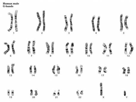
46,XY
The karyotype from a normal male individual comprises 46 chromosomes with one X and one Y chromosome (46,XY), while females have two X chromosomes (46,XX).
IFISH analysis
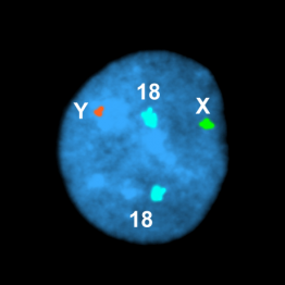
Normal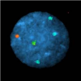
Trisomy 18
Nuclei were hybridized to a red probe for the Y chromosome, a green probe for the X chromosome, and a blue probe for chromosome 18. Nucleus from a normal male, and from a male with trisomy 18.
Chromosome Microarray Analysis
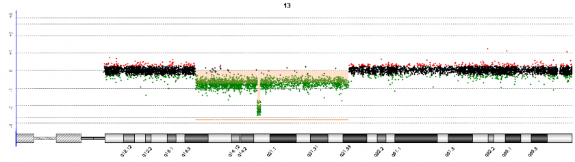
Chromosome 13 deletion
DNA from chronic lymphocytic leukemia cells hybridized to an array. A large deletion of part of chromosome 13 is marked in green and flanked with arrows. Note that a small bi-allelic deletion is also present (open arrow). A schematic of chromosome 13 is shown at the bottom.
Testing Types
- Constitutional Testing: to detect constitutional cytogenetic and genomic anomalies
- Neoplasia Testing: to detect acquired cytogenetic and genomic anomalies in cancer
- Research Testing: to detect cytogenetic and genomic anomalies in research specimens
News: Recommendations for Testing of Fetal Tissue or Products of Conception
Diagnosing a genetic abnormality as the cause of miscarriage allows for recurrence risk counseling, reduces parental feelings of guilt, and eliminates the need for further workup. Unfortunately, maternal cell contamination and culture failure both complicate testing done by traditional chromosome analysis.
Based on a review of the medical literature, existing professional society practice guidelines, chromosome abnormality detection rates using different test methods, and the cost and insurance provider landscapes, the Clinical Cytogenomics Laboratory has developed new recommendations for genetic testing of fetal tissue and products of conception. This tiered testing approach takes advantage of cytogenomic microarray analysis (CMA) and interphase fluorescence in situ hybridization (IFISH), methods which have advantages over karyotyping. Neither requires living cells, producing a higher success rate of obtaining results. Neither requires culturing, allowing detection of monosomies and multiple aneuploidies that aren't viable in culture. And results are thus less susceptible to maternal cell contamination and culturing artifacts. CMA has a further advantage in that it detects subchromosomal alterations not visible with karyotyping.
References